Biopsies

Medical Indications for Skin Biopsies


Different Types of Biopsy Procedures
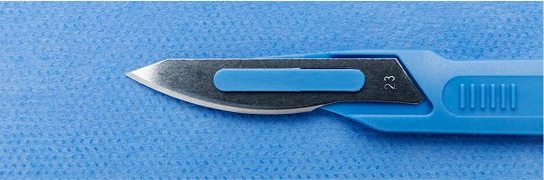
Shave Biopsy
A shave biopsy is performed by removing a thin, top layer of tissue from a lesion or mole using a small blade such as a scalpel or razor. This procedure is typically chosen for the examination of superficial skin abnormalities that may present as suspicious but do not require removal.
Dr. Greenberg will administer a local anesthetic to the affected area before carefully shaving a small portion of tissue for laboratory analysis. This method is effective for diagnosing certain types of skin cancer such as basal cell carcinoma and squamous cell carcinoma. A shave biopsy may also be used for diagnosing abnormal moles on the skin.
This type of biopsy is quick, relatively painless, and poses very little risk of scarring.

Punch Biopsy
A punch biopsy is performed using a special cylindrical blade that penetrates the epidermis, dermis, and, if necessary, subcutaneous fat, to remove a core of skin from the affected area.
Dr. Greenberg may employ this biopsy technique as a means for diagnosing skin rash, infection, and certain types of skin cancer because the amount of tissue extracted can provide a larger sample size for analysis.
This biopsy technique is performed using local anesthesia administered to the affected area prior to the procedure. The amount of tissue removed can vary from 2mm to 5mm in diameter and may require a minimal number of stitches based on the size of the biopsy. In some cases, the wound may be left to heal on its own without suture intervention.
Excisional Biopsy
An excisional biopsy is a technique in which the entire lesion or suspicious, abnormal affected area of the skin is removed for diagnostic analysis using a scalpel. This procedure can be very effective for diagnosing many types of skin cancer including melanoma, and may be employed as a solution for removing the affected tissue in full to prevent the spread of cancerous cells.
Dr. Greenberg will administer a local anesthetic to the area prior to extracting the affected tissue in addition to an appropriate amount of healthy tissue. The reason for using this technique is to gather a sufficient amount of tissue for running a detailed analysis to reach an accurate diagnosis of the affected area.
Once the procedure is finished, the wound is typically closed using stitches. While this type of biopsy is more invasive than other options, the results can provide a higher level of diagnostic detail. There is an increased risk of scarring and post-operative care is necessary in many cases.
Incisional Biopsy
By comparison, an incisional biopsy only removes a portion of the suspicious or abnormal tissue from the affected site and that portion is used for diagnostic analysis. This procedure is often used if the lesion is too large to be removed in its entirety or in cases where removal may present cosmetic or functional concerns.
As with other biopsy procedures, this technique is performed using a local anesthetic applied to the incision site and Dr. Greenberg will remove a portion of tissue that includes both suspicious and healthy samples. Once complete, the area may require minimal sutures or it may be left to heal naturally based on the size and incision site of the biopsy.
In some instances, once a diagnosis is obtained, an additional excision may be required to remove affected tissue.